
Subscribe to our Newsletter



Model No: QH3311-1-2
Advanced PVC human urinary system model for teaching model
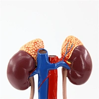
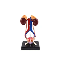
Slightly larger than life size and depicted
in brilliant color, this model of the full
urinary system can easily be seen by
every students in the class. The vena
cava and abdominal aorta are shown; the
right kidney is partially dissected to show
the cortex, medulla, pyrmids, calyces,
pelvis and origin of the renal artery and
vein, and tubing represents the ureters
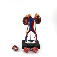
leading to the two-part bladder resting on
the public symphsis. The bladder can be
opened to reveal the mucosa, trigone,
prostatic portion of the urethra, seminal
vesicles, ejaculatory ducts, and vas
deferens. The three piece model is
mounted on a base.
in brilliant color, this model of the full
urinary system can easily be seen by
every students in the class. The vena
cava and abdominal aorta are shown; the
right kidney is partially dissected to show
the cortex, medulla, pyrmids, calyces,
pelvis and origin of the renal artery and
vein, and tubing represents the ureters
leading to the two-part bladder resting on
the public symphsis. The bladder can be
opened to reveal the mucosa, trigone,
prostatic portion of the urethra, seminal
vesicles, ejaculatory ducts, and vas
deferens. The three piece model is
mounted on a base.
Quantity :


Related Products


